Hospital Harnesses Power Of Science, Technology to Improve Surgery for Kids

Photo: Children's National Health System
On the sixth floor of Children’s National Health System in Washington, D.C., engineers, scientists and clinicians develop technologies to make pediatric surgery more precise, less invasive and pain-free. They make up the Sheikh Zayed Institute for Pediatric Surgical Innovation, one of the few research institutes in the country housed directly in a children’s hospital.
“Being physically located within the hospital enables daily communication between multiple specialties,” said Dr. Kevin Cleary, technical director of bioengineering at the institute. “Our scientists and engineers are able to shadow physicians within the medical environment to ensure that solutions developed are clinically relevant. Similarly, physician innovators can bounce an idea by the engineers to assess initial feasibility and collaborate on validated concepts.”
Recently, NIBIB staff had the opportunity to tour the unique institute and get a behind-the-scenes look at some of the projects currently under way, many of which are NIH-funded.
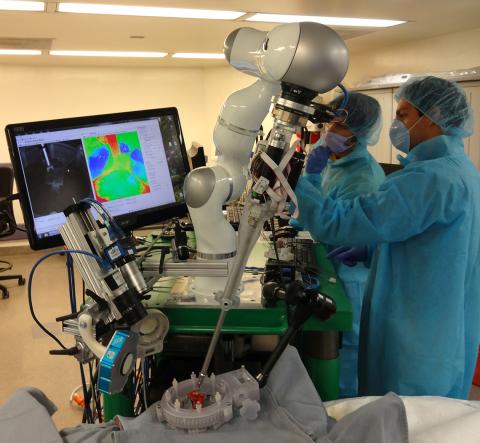
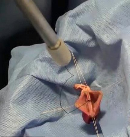
One example of a successful collaboration between an engineer and a surgeon was the development of a surgical robot that can suture tissues all by itself. While robots are commonplace in today’s operating rooms, their movements are almost always directed by a surgeon. In contrast, this robot uses special cameras to track tissue as it shifts or bends while being stitched so that the robot can proceed autonomously.

Photo: Children's National Health System
The researchers’ current goal is to automate a common but complex and delicate task called anastomosis, which is the stitching together of tubular structures in the body, such as arteries or intestines. Dr. Axel Krieger, lead engineer on the project, says the robot can make smaller, more precise stitches than surgeons are able to, lessening the likelihood of leakages, which are a common complication of anastomoses.
The idea for the robot came from Dr. Peter Kim, vice president of the institute and a pediatric thoracic surgeon in the hospital. Kim, who specializes in minimally invasive surgeries, believes a suturing robot could be particularly helpful for use in children.
“Many pediatric surgical procedures require precision, gentleness, maneuverability and fine dexterity,” he said. “Having precise and intelligent tools—including robots that can provide better vision, intelligence and cognition—would be highly beneficial.”

Photo: Children's National Health System
He added that robots in the surgical suite could standardize and improve care for the pediatric population at large by making the best surgical techniques available to all children.
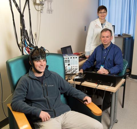
Other exciting projects included a strategy for treating painful bone tumors using magnetic resonance-guided high-intensity focused ultrasound; an imaging headset that could help doctors determine the extent to which very young children are in pain based on activity patterns in their brain; and software for planning difficult surgeries on children with skull abnormalities.
Throughout the tour, researchers stressed many of the unique challenges associated with developing surgical technologies and devices for children. For example, pediatric devices often stay in the body longer and, therefore, need to be more durable. Devices also need to adapt as children grow to avoid repeat surgeries. In addition, a number of surgical robots are being developed that would enable surgeons to operate on children inside an MRI scanner. This would lessen their exposure to radiation generated by CT and fluoroscopy, imaging modalities currently used by surgeons to visualize tissues during an operation.
“What struck me was the breadth of technologies being developed,” said Dr. Steven Krosnick, a program director at NIBIB. “To me, it demonstrates the power of bringing together clinicians, engineers and scientists to solve complex challenges in medicine. Surgeons often have a sense that if only they had a specific tool or imaging technology, they could do their job better, faster, safer. The problem is they don’t generally have the time or the expertise to develop it. It’s the collaboration with the engineers and scientists that can turn those ideas into reality.”
