BRAIN Initiative Contest Garners Eye-Catching Photos, Videos

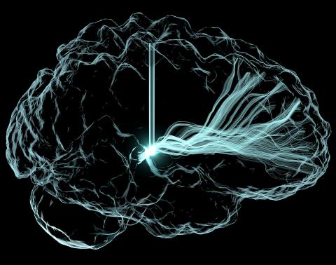
Photo: Andrew Janson
Neuroscience imagery has come a long way since the hand-drawings of Spanish scientist-artist Santiago Ramón y Cajal. Yet, images, and now videos, continue to capture the wonder and beauty natural to the exploration of the brain.
NIH recently hosted its first-ever BRAIN Initiative “Show Us Your Brains!” Photo and Video Contest as a fun way to showcase and demonstrate to the public the creativity and artwork of scientists working on the BRAIN Initiative.
The contest was held as part of the 5th annual BRAIN Initiative Investigators Meeting Apr. 11-13 at Washington Marriott Wardman Park Hotel in Washington, D.C. Cool, eye-catching image submissions were encouraged from anyone engaged in the initiative, regardless of discipline, career stage or funding source.
The program committee reviewed a total of 58 submissions—36 photos and 22 videos—and selected entries they felt best captured the creative spirit of the initiative. Those images then were posted online for public voting; the winning top 3 photos and top 3 videos were announced Apr. 13.
Contest winners are listed below. To view the full group of winning images, visit https://bit.ly/2Un2xnX.
Photo Winners:
- First Place: “Light Me Up!” from Andrew Janson, graduate student research assistant, Scientific Computing and Imaging Institute, University of Utah. Light-based rendering of deep brain stimulation’s electrical excitation of neuronal fiber pathways to treat patients with traumatic brain injury.
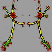
- Second Place: “Dancing Devils” from Dr. Sharada Tilve, postdoctoral fellow, Geller Lab Group, NHLBI. Mouse hippocampal neuron stained for f-actin (red) and tubulin (green) look like the devil dancing ballet.
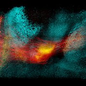
- Third Place: “Neural circuit in the storm” from Dr. Young-Gyun Park, postdoctoral fellow, Chung Lab, MIT. 3-D image of parvalbumin+ neurons (red, neurites; green, presynaptic puncta) swimming through the waves of GAD1+ (cyan) neurons.
Video Winners:
- First Place: “High-Resolution MORF3-labeled Hippocampal Neurons” from Dr. X. William Yang, UCLA and Dr. Kwanghun Chung, MIT. Using MORF3 and SHIELD, pyramidal neurons were sparsely labeled and imaged at very high resolution deep within a whole hemisphere.
- Second Place: “3-D Diffusion Tractography” from James Stanis, medical animator, Mark and Mary Stevens Neuroimaging and Informatics Institute, USC.
- Third Place: “Neural circuit in the storm,” which placed in both categories.


