Congressional Staff Discover Innovative NIBIB Biotech

Photo: Marleen Van Den Neste
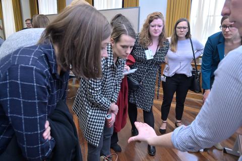
A smartphone app to help make health care more accessible for people with anemia who need to measure their hemoglobin levels. An optical imaging system to see nerves under the skin during surgeries. These were two of the technologies recently showcased by NIBIB scientists and grantees to a group of 40 congressional staffers. The recent congressional tour at NIH exhibited National Institute of Biomedical Imaging and Bioengineering-supported biotechnologies and gave the visitors a hands-on experience with a broad spectrum of research projects.
NIBIB director Dr. Bruce Tromberg opened the day with brief remarks highlighting NIBIB’s history and impact at NIH. He joined the five groups of exhibitors in demonstrating to congressional staff the innovative biotechnologies that address health problems.
“The event was a great success,” said Tromberg. “NIBIB takes its charge seriously about informing the public and Congress on how taxpayer dollars are spent. These projects are wonderful examples of how NIBIB-funded research leads to the development of game-changing technologies that benefit patients.”
The tour came at the invitation of the American Institute for Medical and Biological Engineering (AIMBE) and was organized in part by NIBIB’s Office of Science Policy and Communications. AIMBE is an advocacy group that supports medical and biological innovation and aims to increase public understanding of scientific advances.

Photo: Marleen Van Den Neste
The presentations included a demonstration from NIBIB’s newest intramural investigator Dr. Kaitlyn Sadtler, chief of the immunoengineering section. She showcased protective gels that help program an immune response to promote the integration and growth of engineered tissues. This work could accelerate the development of next-generation materials used in medical devices such as pacemakers and drug delivery vehicles.
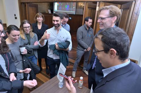
Dr. Wilbur Lam and his postdoctoral fellow Dr. Robert Mannino from Emory University and Georgia Institute of Technology presented their smartphone app that non-invasively measures hemoglobin levels in patients with anemia, the most common blood condition. Hemoglobin levels are currently measured through blood tests. By using a picture of a person’s fingernails, accurate hemoglobin levels can be obtained without a trip to the clinic. The new technology could be useful in settings where medical resources are limited.
Dr. Bin He, a professor at Carnegie Mellon University, and his postdoctoral fellow Dr. Abbas Sohrabpur, explained their brain-mapping technique that helps surgeons plan for operations on patients with epilepsy. The imaging technique uses novel machine-learning algorithms and EEG test results taken during a patient’s seizure. The resulting images show physicians the sites where seizures are originating and help them understand the disease better.

Photo: Marleen Van Den Neste
Congressional staff handled and examined 3-D printed phantom tissues created at the University of Maryland by Dr. John Fisher and his team. Graduate student Sarah Van Belleghem and Dr. Bhushan Mahadik gave presentations on how the biomaterials can help build scaffolds to repair damaged tissues or deliver drugs to precise locations. Dr. Guang Yang showcased a 3-D printed bioreactor that helps supply cells or tissues with nutrients in a controlled manner, so cell or tissue interactions can be more easily studied.
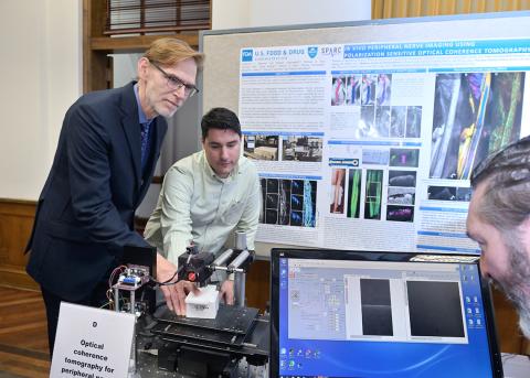
Legislative aides had the opportunity to look under their skin and see their nerves with an imaging technology brought by a team of researchers from the FDA and Massachusetts General Hospital. FDA scientists Drs. Srikanth Vasudevan and Daniel Hammer guided and imaged the hands of aides using the advanced optical imaging system. The system may provide better outcomes during operations by helping surgeons visualize nerves below the skin.
Visitors also viewed apps created by NIBIB’s Office of Science Policy and Communications. The Understanding Medical Scans app uses images and videos to provide patients with information about the various types of medical scans many people undergo during an illness. The team also showcased their Surgery of the Future app that displays what a futuristic surgery room may look like with the help of NIBIB-supported technologies. Both apps are available for free download on most devices.
