NIH-Funded Tissue Chips Push New Boundaries in Space

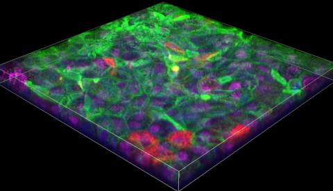
Photo: Massachusetts Institute of Technology
With the help of a Falcon 9 rocket, NIH-supported research teams sent tiny, bioengineered models of human organs hurtling toward the International Space Station U.S. National Laboratory (ISS National Lab) at thousands of miles per hour.
The launch, which carried tissue chips modeling heart and gut tissue, took place Mar. 6 from Cape Canaveral, Fla.
Tissue chips are miniature, 3-D models of human tissues such as the lung and liver, designed to mimic functions of the human body and support living human tissues and cells.
By studying heart and gut tissue models on the ISS National Lab, researchers hope to learn more about molecular changes in heart tissues exposed to the extreme environment of microgravity and get new insights into immune responses in the intestine that could help improve human health back on Earth.
The low-gravity environment on the ISS National Lab presents valuable opportunities for research that are not found on Earth. For example, low-gravity environments can cause changes in the human body that are similar to accelerated disease and aging processes.
Because these changes happen relatively quickly, the tissue chips sent to the ISS National Lab allow researchers to model and study—on a much shorter timescale—conditions related to aging and disease that might take years to develop on Earth.
Both projects are funded through the Tissue Chips in Space initiative, which is a collaboration among the ISS National Lab, NCATS and NIBIB.
