On the Cover
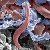
Scanning electron micrograph of a sickled red blood cell (red) inside the circulatory structures of the human spleen, whose function is to filter and remove unhealthy red blood cells, including rigid cells such as sickle-shaped cells. The blue cells indicate the wall of a splenic sinus (small vein where red blood cells are filtered). The gray structures indicate the splenic cords—cellular clusters where the red blood cells are checked by macrophages for the presence of surface alterations or certain antibodies. The image was produced by NHLBI-funded scientist Dr. Pierre Buffet of Inserm-University of Paris, whose research explores the role of the spleen in sickle cell disease. September is Sickle Cell Disease Awareness Month.
Pierre Buffet/Inserm-University of Paris


