Wall of Watercolors
2D Microscopes Featured in CC Gallery

Photo: lillian fitzgerald
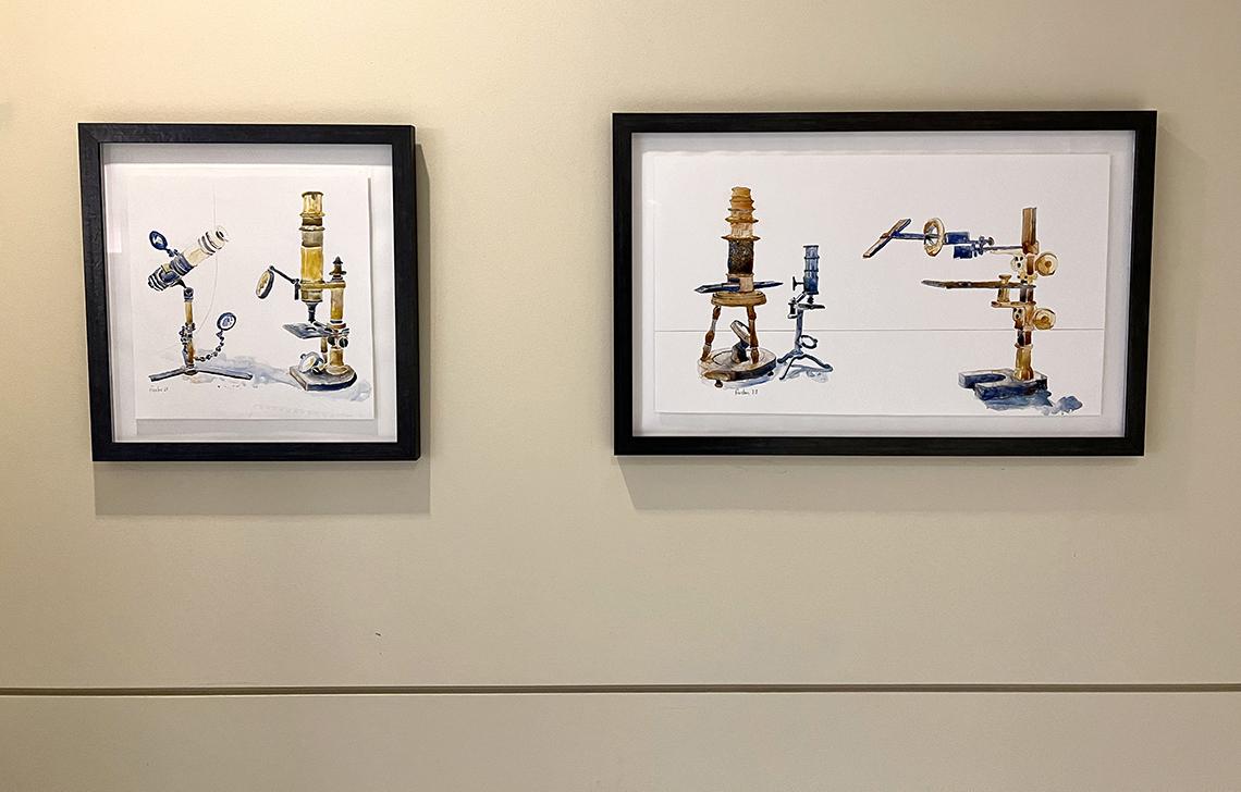
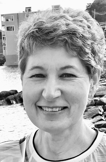
Scientists and other research enthusiasts at NIH can view medical instruments in a novel way: A wall of watercolors featuring microscopes painted as portraits by artist Sue Fierston was recently installed at the Clinical Center.
“I love thinking about these microscopes and instruments as beings from another place and time,” said Fierston. “They carry a cultural record with them: the round wooden German microscope here was fashioned by German toymakers who were used to carving wood with a delicate hand.”
Microscopes, before electricity and the lightbulb, were designed to capture as much light as possible, she explained. “Microscope designers tried everything to illuminate the stage of the microscope including candles, bullseye lenses and mirrors. These microscopes had fanciful, almost animated shapes as designers aimed lenses or lengthened tubes in an attempt to see the magnified objects.”

Photo: lillian fitzgerald
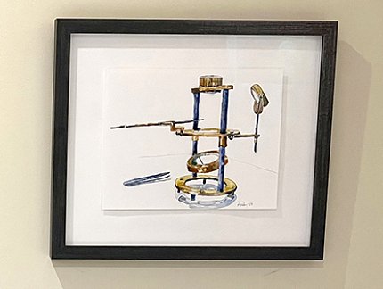
Fierston drew the microscopes on site at the University of California, Berkeley, in October 2022. Dr. Steven Ruzin, curator of the historic Golub Collection, allowed her to choose any microscopes that caught her attention.
“I sat with them and positioned them as I wished and then drew them from life,” the artist recalled. “I took photos at the time to help me remember the lighting and reflections because I did my painting back home in Maryland.”
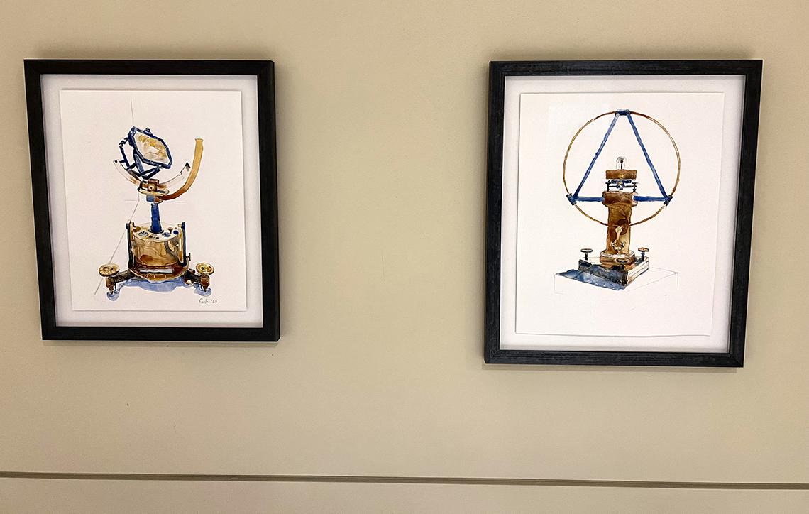
The two historic scientific instruments, the tangent galvanometer and the heliostat, come from the collection of Mark McElyea. “I drew them at an exhibit of his collection that appeared at the San Francisco International Airport in 2022,” Fierston said. “I found them by chance as I was traveling through.”
The series is a continuation of one she began around 2007. At that time, the artist painted eight historic microscopes from the Billings Collection at the National Museum of Health and Medicine (NMHM). The work was shown at NIH, NMHM and the Lombardi Cancer Center in Washington, D.C.

“My research and painting took about a year from initial idea to framed paintings,” Fierston said. “At one time I hoped to paint them in colorful acrylics on pizza boxes! My idea was to offset their seriousness with a fanciful painting style. But I faced a deadline and, in the end, I returned to watercolor on the plastic surface Yupo with the limited color palette of French ultramarine/cobalt and burnt/raw sienna.
“I also love the challenge of painting reflections on silver and brass,” she continued. “Yupo gave me an opportunity to do this abstractly, because watercolor runs randomly across the plastic until the pigment dries. As an artist, I am drawn to the contrast between a carefully drawn microscope and the random paint that fills the image.”
In 2022, Fierston was awarded an artist grant from the Arts and Humanities Council of Montgomery County to help support the project. The funding enabled her to frame the paintings and pay for supplies.
Fierston’s exhibition is on display until Jan. 5, 2024 in the first floor corridor of the Clinical Center.

Photo: lillian fitzgerald
