


On the Cover
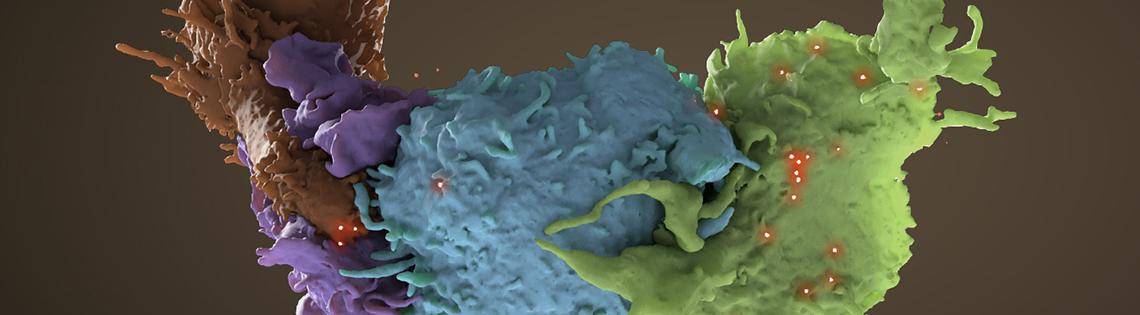
3D structure of HIV infected (blue, green) and uninfected (brown, purple) T cells interacting. One cell (brown) has wrapped an extension around its uninfected neighbor (purple) to reach an infected cell (blue). Data comes from focused ion beam scanning electron microscopy.
donny bliss, nlm; sriram Subramaniam, nci