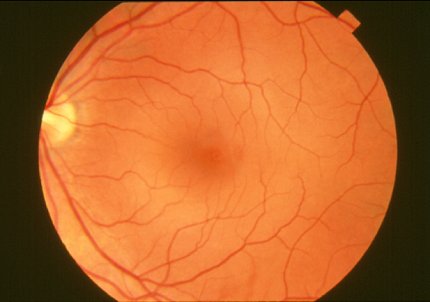
NIH Scientists Combine Technologies to View the Retina in Unprecedented Detail

Photo: NEI
By combining two imaging modalities—adaptive optics and angiography—investigators at the National Eye Institute can see live neurons, epithelial cells and blood vessels deep in the eye’s light-sensing retina. Resolving these tissues and cells in the outermost region of the retina in such unprecedented detail promises to transform the detection and treatment of diseases such as age-related macular degeneration, a leading cause of blindness among the elderly. The paper was published online in Communications Biology.
“For studying diseases, there’s no substitute for watching live cells interact,” said Dr. Johnny Tam, Stadtman investigator in NEI’s clinical and translational imaging unit and lead author of the paper. “However, conventional technologies are limited in their ability to show such detail.”
Biopsied and postmortem tissues are commonly used to study disease at the cellular level, but they are less than ideal for watching subtle changes that occur as a disease progresses over time. Technologies for noninvasively imaging retinal tissues are hampered by distortions to light as it passes through the cornea, lens and the gel-like vitreous in the center of the eye.
Tam and his team turned to adaptive optics to address this distortion problem. The technique improves the resolution of optical systems by using deformable mirrors and computer-driven algorithms to compensate for light distortions. Widely utilized in large ground-based space telescopes to correct distortions to light traveling through the atmosphere, adaptive optics began being used in ophthalmology in the mid-1990s.
The NEI researchers combined adaptive optics with indocyanine green angiography, an imaging technique commonly used in eye clinics that uses an injectable dye and cameras to show vessel structures and the movement of fluid within those structures.
