New Non-Invasive Imaging Tool Maps Labor Contractions

Photo: Washington University in St. Louis
NIH-funded researchers have developed a new imaging tool, called electromyometrial imaging (EMMI), to create real-time, three-dimensional images and maps of uterine contractions during labor. The non-invasive imaging technique generates new types of images and metrics that can help quantify contraction patterns, providing foundational knowledge to improve labor management, particularly for preterm birth.
The tool lays the foundation toward potentially predicting who is at risk to deliver prematurely or whose labor pattern might result in the need for a cesarean section delivery.
The study team initially developed EMMI using a sheep model. In the new study, the team tailored EMMI for human clinical use and tested it among a group of 10 women with healthy pregnancies. Current clinical methods to measure contractions (i.e., tocodynamometry and an intra-uterine pressure catheter) can provide only limited details, such as contraction duration and intensity, while also being invasive.
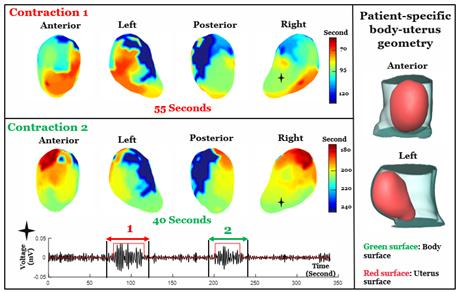
EMMI integrated two types of non-invasive scans—a fast anatomical MRI to obtain an image of the uterus and a multi-channel surface scanning electromyogram that uses sensors placed along the belly to measure contractions during labor.
These data are then combined and processed into three-dimensional uterine maps, with warm colors denoting areas of the uterus that are activated earlier in a contraction, cool colors indicating areas that are activated later and gray areas showing inactive regions. A sequence of maps is generated over time, creating a visual time lapse that shows where contractions start, how they spread and/or synchronize, and potential patterns that are associated with a typical pregnancy versus one with complications.
Results from the pilot study also bring clarity to a longstanding question on how contractions begin—EMMI data suggest there is no fixed, pacemaker-like region in the uterus that initiates labor.
EMMI offers new possibilities for better understanding human labor and facilitating development of optimized, patient-specific interventions. The authors note that an EMMI contraction atlas generated from healthy pregnancies can serve as a resource to understand and diagnose preterm labor and possibly identify patients who would benefit from an induction versus those who may need a cesarean section.
The small study is supported in part by NICHD through its Human Placenta Project and other programs. Findings are published in Nature Communications.
