On the Cover
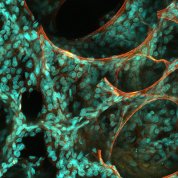
Human pediatric lung alveolar walls (green-stained nuclei) draped over elastin fibers (red), as seen through a multi-photon microscope. An individual alveolus, the gas-exchanging structure of the lung, is about the thickness of a sheet of paper. The image is part of NHLBI’s LungMAP project, a historic effort to understand the molecular and cellular architecture of the human lung. This year marks the 50th anniversary of the founding of NHLBI’s Division of Lung Diseases. November is National COPD Awareness Month.
Gloria Pryhuber, Cory Poole, University of Rochester Medical Center, supported by NHLBI


