NIEHS Cryo-EM Resources Support Fight Against Novel Coronavirus

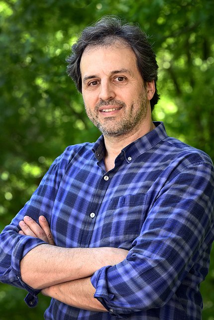
Photo: Steve McCaw
The NIEHS cryo-electron microscopy (cryo-EM) facility, led by Dr. Mario Borgnia, is providing key support to the Duke Human Vaccine Institute (DHVI) in the fight against the SARS-CoV-2 virus, which produces COVID-19. Recently, Borgnia spoke with NIEHS about the research he conducts with Duke’s Dr. Priyamvada Acharya.
Cryo-EM is an advanced microscopy platform launched at NIEHS in 2017 as part of the Molecular Microscopy Consortium, along with Duke and the University of North Carolina at Chapel Hill.
“I am so glad we invested in cryo-EM technology,” said NIEHS scientific director Dr. Darryl Zeldin. “Mario is doing an outstanding job leading the Molecular Microscopy Consortium, to provide support for the entire region. Our investment is paying off as Mario is working collaboratively with scientists at DHVI to facilitate development of a vaccine against SARS-CoV-2.”
Why are you focusing on the so-called spikes of the virus structure?
Borgnia: The spikes that form the so-called corona are viral proteins. Members of the coronavirus family bud out new viral particles from an infected cell by pinching a small bubble of the cell’s own membrane.
This envelope surrounds the virus’ genetic material, acting as a cloak to prevent detection. The body’s immune system does not recognize the virus as foreign, so it does not mount a fight. Yet the virus at this point is still isolated in its own bubble.
Here is where the spike comes into play. If you think of a key and lock, the spike is the key. The lock is a receptor in the human cell. The virus attaches the key in a new cell’s lock. It then fuses its envelope with the cell membrane and injects its genetic material into the cell.
But the spikes are also the Achilles heel of the virus, because the immune system can recognize them as foreign material.
During the early stages of viral infection, the body begins generating antibodies against the spikes, or any portion it recognizes as foreign. If it does this faster than the virus replicates in the body, we do not get really sick. The idea of a vaccine is to prime the immune system with the spike protein to increase the concentration of antibodies against it, even before the body detects a live virus.
Once our immune system knows the disease, it has the advantage and can drive the virus away. The goal of our work is to generate a version of the spike that prompts the body to generate effective antibodies.
This is very different from HIV, for example, which is much more complicated. HIV mutates in the body so that infected people rarely develop protective immunity, although we are learning tricks to teach the immune system to fight HIV as well.
A major goal in the effort to defeat this [coronavirus] pandemic is finding a way to interfere with the process of cellular infection. A treatment would block the virus’s recognition of the target receptor in those who are sick. A vaccine would teach the immune system to make antibodies to neutralize the spikes before disease develops.
Using cryo-EM, we hope to determine the structure of the spike—by itself, in complex with the target receptor, and in complex with neutralizing antibodies.
Where in the process are you right now?
Borgnia: Dr. Acharya’s team is working closely with Allen Hsu, here at NIEHS, to optimize cryo-EM grids for SARS-CoV-2 spike samples using the NIEHS Talos Arctica microscope. These are then imaged using the Duke Titan Krios microscope. Dr. Acharya’s group is working around the clock together with my team to further optimize the specimens.
Can you explain what optimizing the specimens involves?
Borgnia: To get a structure using cryo-EM, you gather tens of thousands of images of the protein, then average them to obtain a 3-D structure. To do this, the proteins are frozen in a thin layer of ice on a grid, by a process known as vitrification.
By optimizing the vitrification conditions, we can produce cryo-EM grids suitable for high-resolution imaging. We look forward to continuing our work with Dr. Acharya’s group to optimize samples of spike variants and complexes for imaging.
