Now showing at Strathmore
‘Microscopy as Masterpiece’ Exhibits NIH-Funded Images
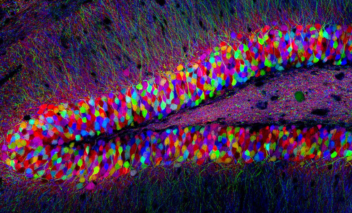
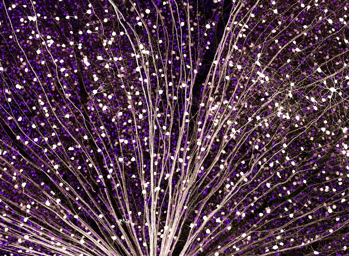
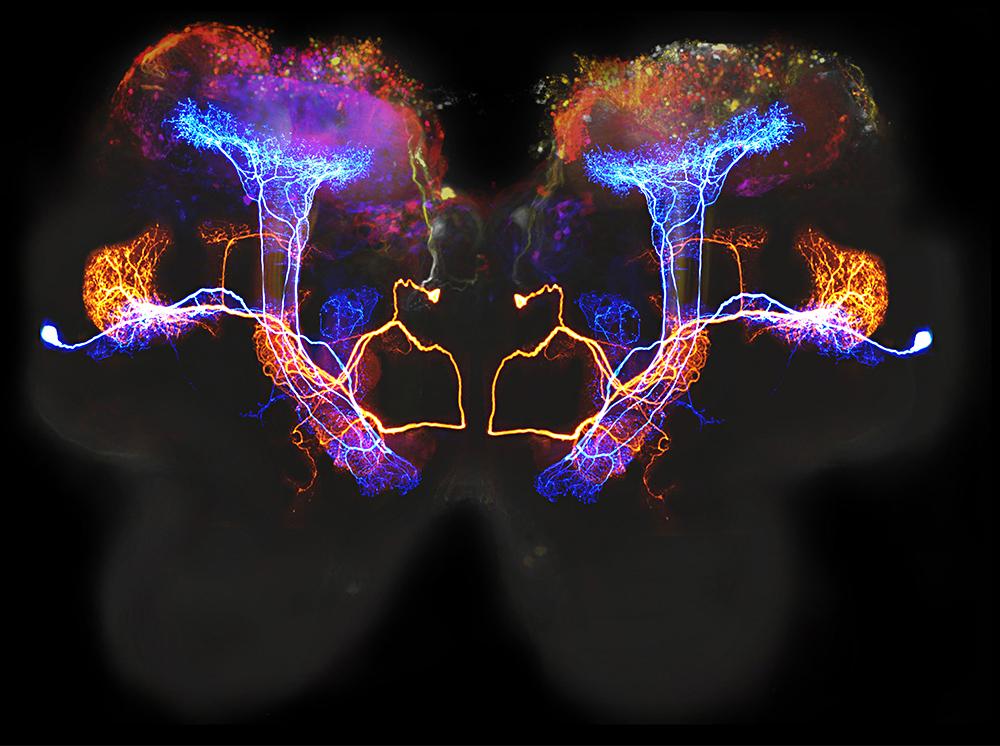
Colorful scientific images and videos from NIH researchers and grantees greet attendees at Strathmore Mansion’s “Arts & the Brain” lecture series. The digital presentation, displayed on a large flatscreen monitor, features nerve cells in jelly bean colors, star-shaped glial cells, a flyover of boutons bursting with synaptic vesicles and much more.
“Attendees have been taken with the beauty of these images,” said Lauren Campbell, director of education at Strathmore and mastermind of the Arts & the Brain program. “The Microscopy as Masterpiece exhibit adds a surprising and delightful visual and concrete element to the lectures.”
Campbell created the lecture series to appeal to the community’s interest in blending art and science. “Many people in Strathmore’s orbit are both science-focused and arts-focused, and have lots of talent in both areas,” she said.
With NIH and Strathmore just one Metro stop apart, a collaboration seemed natural. The digital exhibit grew out of a conversation Campbell had with a musician who performed at the mansion—and also happened to be an NIH employee.
The final lecture in the 2017 Arts & the Brain series, titled “Medical Avatar,” will be on Thursday, June 1. For information, see https://www.strathmore.org/education/programs-for-adults/arts-the-brain-package.
Tickets to the lecture cost $25. Viewing the Microscopy as Masterpiece exhibit before or after each lecture is free.


