Competing Neurons
Neuroscientist Studies the Making of Memories

Photo: Marleen Van Den Neste
We’ve all had that light-bulb moment, suddenly recalling a memory, not even sure how we plucked it out of its latent state. Behind the scenes, there were all kinds of neurons, molecules and synapses at work.
Dr. Sheena Josselyn, senior scientist at the Hospital for Sick Children in Toronto, has long studied what triggers the brain to make, retrieve and link memories. It’s a field that provides insights into how we learn and could lead to interventions for cognitive impairments and neurodegenerative diseases.
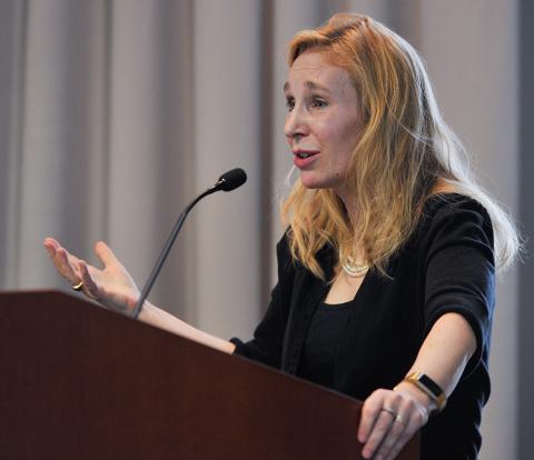
“My lab is really interested in understanding how the brain encodes, stores and uses information,” said Josselyn, who spoke at a recent NIMH Director’s Innovation Speaker Series lecture at the Neuroscience Center. “We think this is a real fundamental goal of neuroscience.”
Building on basic research, Josselyn’s lab began searching for the long-elusive engram, or memory trace—a change in the brain when something is encoded as a memory. Previous researchers searched for it for years but couldn’t pinpoint the engram to a specific spot in the brain.
To begin her quest, Josselyn ran a simple Pavlovian conditioning experiment on mice. After a quick shock followed by a tone, she placed the mouse in a different setting and played the tone again. The moment the mouse froze to the tone would indicate memory formation.
Research has shown that neurons in the lateral amygdala are involved in the memory trace. “They respond both to the tone and to the shock,” said Josselyn. “Yet there’s a surprisingly small number, about 20 percent, that seem to be necessary to encode any one given fear memory.”
In humans, fear memories are sparsely encoded in the amygdala, leading Josselyn to question why certain neurons are chosen to be an engram team player while others get excluded. Her experiments revealed the highly competitive nature of neurons.

Photo: Marleen Van Den Neste
“Eligible neurons seem to compete against one another,” said Josselyn. “The ones that win this competition and actually become part of this fear memory engram are the ones with the highest level of transcription factor CREB [cAMP response element binding protein].”
It turns out that CREB holds important clues to memory formation in part because it can increase neuronal excitability. These enthusiastic neurons are more likely to get recruited into the engram.
When CREB was increased in a small portion of random neurons, the mice froze more often, indicating better memory. Josselyn’s lab then overexpressed CREB in select neurons and, after the mouse learned something, shone a light to turn off those cells overexpressing CREB; these mice showed a memory deficit.
“We were very excited about this,” she said. “It was the first hint we’d gotten our hands, or at least our viruses, on the engram…These findings lead us to conclude that lateral amygdala neurons overexpressing CREB at the time of training are somehow necessary, or indispensable, for the subsequent expression of that fear memory.”
Further experiments elucidated how some memories might be linked. In one study, Josselyn found that if two fear memory events occurred within 6 hours of each other, they were allocated to the same population of neurons, which linked the memories. Other researchers are currently studying this phenomenon in people, using functional MRI.
“They’re looking at fMRI codes in broad brain regions,” Josselyn said. “We’re looking at specific populations of a small brain region, but the goal is to link up these human and these mouse studies.”
Josselyn is also studying what might cause memory disruptions in Alzheimer’s disease. In recent experiments, she injected mice with amyloid beta, peptides that can induce neuron degeneration and disrupt synapses, which are prevalent in the brains of Alzheimer’s patients. She then injected the transgenic mice with a peptide that would restore the synapse, which returned memory in these mice. Other researchers have made similar strides with various molecular interventions in animal models, she noted, but to date none have succeeded in people.
In these experiments, Josselyn also had another curious finding. In wild type mice, the neural freezing signature, or memory recall, started with individual cells, then evolved into a coordinated network activity. This pattern completion scheme was disrupted in the transgenic mice but was restored after the intervention.
“We think that events are represented by patterns of activity in cells that are active during an event, an engram, and we think memory retrieval involves recapitulation of this pattern of sequences.”
Josselyn said it’s time to take what researchers have learned so far about genes and circuit dynamics and use computational tools to look at all aspects of behavior.
In people, recalling a memory is an active process that could change the underlying engram. “You can change a memory [every time] you recall it,” said Josselyn during the post-lecture discussion. “You can extinguish it. You can strengthen it. You can pair it with something new. When you change it, it becomes plastic again.”
One attendee asked about the possibility of manipulating traumatic memories.
“When we retrieve an older memory, the neurons important in the recovery of this memory are active,” she said, “and I think that gives us a really cool time window to try an intervention.”
