Life Magnified
NIH Zebrafish Research Featured on Postage Stamps
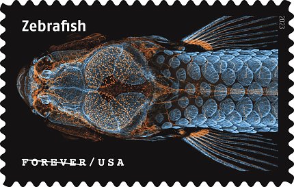
Photo: U.S. Postal Service
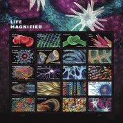
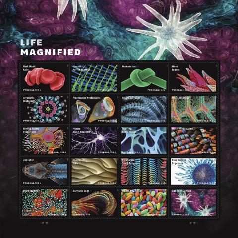
A microscopy image created by NIH researchers is part of the “Life Magnified” stamp panel issued Aug. 10 by the United States Postal Service (USPS).
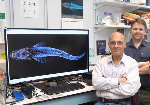
The NIH zebrafish image, which was taken to understand lymphatic vessel development in the brain, merges 350 individual images to reveal a juvenile zebrafish with a fluorescently tagged skull, scales and lymphatic system.
“Zebrafish are used as a model for typical and atypical human development. It is surprising how much we have in common with zebrafish,” said Dr. Diana Bianchi, director of the Eunice Kennedy Shriver National Institute of Child Health and Human Development (NICHD), which generated the image. “NIH research affects our lives every day. My hope is that this postage stamp will help spur conversations and appreciation for the importance of basic science research.”
The image was taken by NICHD’s Daniel Castranova, an aquatic research specialist, with assistance from former trainee Bakary Samasa. The research was conducted in the section on vertebrate organogenesis, led by principal investigator Dr. Brant Weinstein. The lab is devoted to understanding mechanisms guiding the formation of blood and lymphatic vessels. The image also received top honors in the 46th annual Nikon Small World Photomicrography Competition in 2020.
Findings from the microscopy image were published in Circulation Research and featured on the journal’s cover. The work led to a groundbreaking discovery that zebrafish have lymphatic vessels inside their skull. These vessels were previously thought to occur only in mammals, and their discovery in fish could expedite and revolutionize research related to treatments for diseases that occur in the human brain, including cancer and Alzheimer’s.
Life Magnified, a set of 20 Forever stamps, also includes work from other researchers relevant to the broader NIH community. Two creators lead microscopy core facilities often used by NIH-funded investigators at their universities. Dr. Tagide deCarvalho, director of the Keith R. Porter Imaging Facility at the University of Maryland, Baltimore County, created “Moss Leaves” and “Mold Spores.” Jason M. Kirk is director of the Optical Imaging & Vital Microscopy Core at Baylor College of Medicine. He created “Oak Leaf Surface” and “Mouse Brain Neurons.”
