NIH Record - National Institutes of Health
A multicolored fluorescence microscopy image. Bright blue dots, interspersed with red, purple, yellow and white lines cover a black surface.

September 26, 2025

  • Balancing a Fluid Situation

    NIH-funded scientists created a 3D map of kidney nerves and vascular components at an unprecedented scale. Understanding kidney structure and function at this level of detail may provide new clues toward future treatments for conditions that affect kidney development or cause kidney-related disease.
A multicolored fluorescence microscopy image. Bright blue dots, interspersed with red, purple, yellow and white lines cover a black surface.

On the Cover

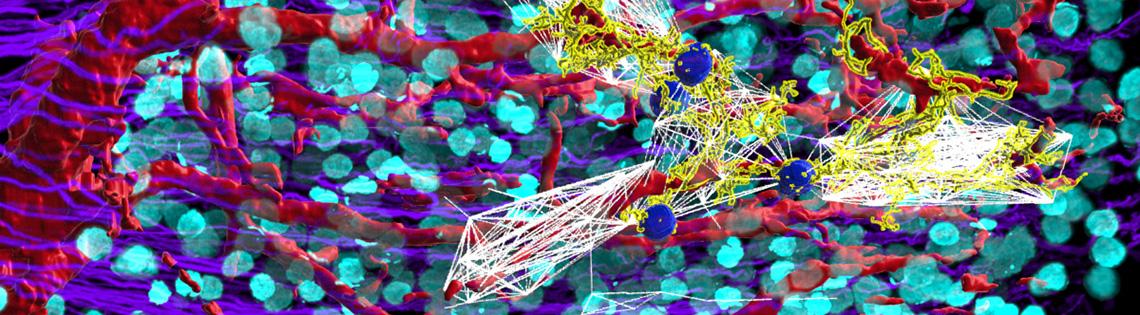
3D 5x light sheet fluorescence microscopy image of adult human kidney cortex. Vessels are red, glomeruli are cyan and collecting ducts are in purple. A selection of the neural network that was isolated among several glomerular communities is shown by the yellow nerves, with the white lines representing glomerulus-to-glomerulus neural connections. At the center, in blue spheres, are ‘mother glomeruli’, which exist as hub points in the overall network.

Liam McLaughlin, Sanjay Jain Lab, Washington University at St. Louis

The NIH Record

The NIH Record, founded in 1949, is the biweekly newsletter for employees of the National Institutes of Health.

Published 25 times each year, it comes out on payday Fridays.

Editor: Dana Talesnik
Dana.Talesnik@nih.gov

Assistant Editor: Eric Bock
Eric.Bock@nih.gov

Assistant Editor: Amber Snyder
Amber.Snyder@nih.gov