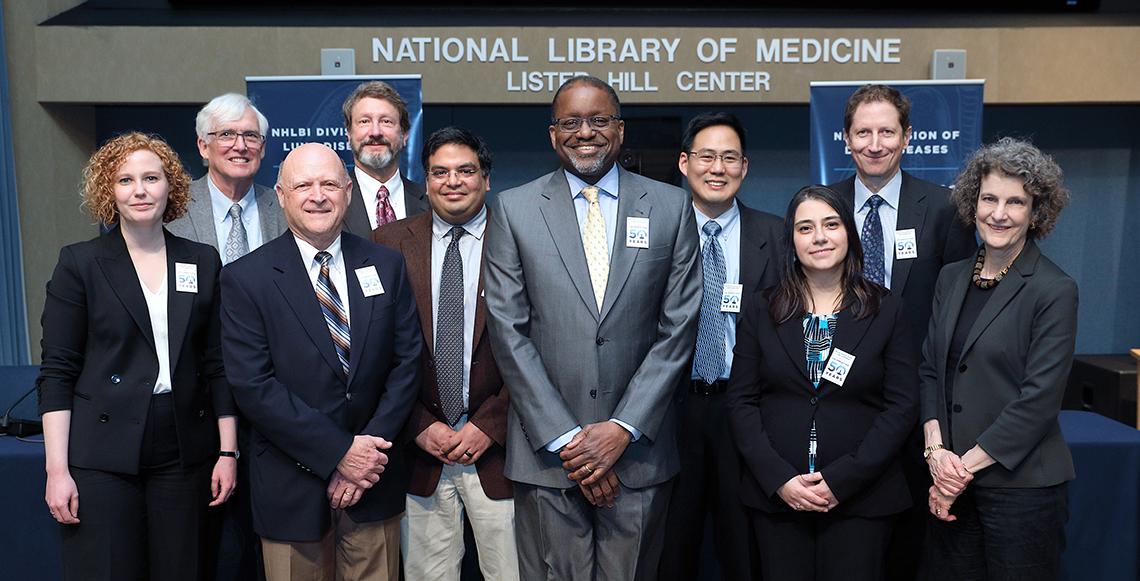
NHLBI Division Celebrates 50th Anniversary

For 50 years, the Division of Lung Diseases, part of the National Heart, Lung, and Blood Institute, has focused on translating basic scientific research into more effective treatments and patient care.
In April, the division kicked off its golden anniversary with a scientific symposium highlighting the progress and future of a centerpiece of this work—lung imaging. The half-day event included researchers from both the intramural and extramural programs. They discussed how they apply sophisticated imaging techniques to advance the understanding of healthy and diseased lungs at the cellular and molecular level and how this work informs the diagnosis, treatment and management of lung diseases in general.
In welcoming remarks, NHLBI director Dr. Gary Gibbons acknowledged the institute’s legacy of excellence in pulmonary science research since 1969 and said he is excited about the breakthroughs yet to come. With technological innovations in imaging, the phrase “chronic lung disease” one day will no longer be needed, he said, noting that he has charged the division leadership with making that a reality sooner than later. With a renewed commitment to the prevention, preemption, remission and reversal of pulmonary disorders, Gibbons said he is sure the watershed moment will come.
Dr. James Crapo of National Jewish Health gave the first talk, highlighting a key finding of the NHLBI-funded COPDGene study he leads as principal investigator. After following a cohort of 10,000 people for 5 years, Crapo said he had identified a group of participants who, after taking a breathing test that measures lung function, showed no obstruction in their airways, even though they still had symptoms of COPD. These participants also had a higher risk of morbidity and mortality, compared to a group who had showed gradual lung tissue damage over time. With this finding, Crapo has a proposal: to include people who have not yet developed airflow obstruction in the diagnosis of COPD as a way to help prevent disease.
Dr. Scott Fraser of the University of Southern California noted his work with the NHLBI-funded Molecular Atlas Lung Development Program consortium. He and his team are building tools that can acquire and analyze microCT images in hopes of defining the anatomy of the developing lung.
Dr. Sanjay Jain of Johns Hopkins University discussed his progress developing molecular imaging tools to better understand bacteria, the infections they cause and the environments that could minimize them. The hope is that this will lead to limiting the overuse of antibiotics.
NHLBI’s Dr. Adrienne Campbell-Washburn showed how advances in low-field MRI could greatly improve cardiac and lung imaging and other image-guided procedures that diagnose and
treat disease.
Kicking off the second session, Dr. Carmen Priolo of Brigham and Women’s Hospital/Harvard Medical School detailed how PET imaging can identify abnormal metabolic processes in preclinical models—and one day in imaging trials—of lymphangioleiomyomatosis (LAM), a rare lung disease mostly affecting women.
NHLBI’s Dr. Marcus Chen illustrated how an ultra-low dose chest CT, when compared to a traditional x-ray, could be used to validate clinical cases of LAM and other lung diseases.
Dr. R. Graham Barr of Columbia University rounded out the symposium by discussing how he uses machine learning and advanced imaging techniques to help understand chronic lung disease in large-scale, long-term studies, including the Multi-Ethnic Study of Atherosclerosis lung study and the Subpopulations and Intermediate Outcome Measures in COPD Study.
Dr. James Kiley, who has directed the Division of Lung Diseases since 1984, ended the event, calling the discussions “a window into the future of pulmonary imaging at all levels. “It was a phenomenal display of hard work by many people across the country,” he said, inviting the audience to stay tuned for more anniversary events to come.
