Stoked by Stochasticity
Betzig Boosts Biology with Better Visuals

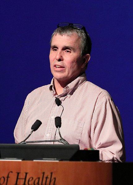
Photo: Chia-Chi Charlie Chang
While there are those who think necessity is the mother of invention, Nobel laureate Dr. Eric Betzig says dissatisfaction—pure, serial frustration with limits—has prompted him to create sophisticated new microscopes that are allowing biologists to understand the lives of cells at more intimate levels.
Convinced that you can’t understand what you can’t see, especially in living systems, Betzig, a physicist with appointments at both the University of California, Berkeley, and the Howard Hughes Medical Institute, shared “The Secret Lives of Cells” at a Wednesday Afternoon Lecture on Apr. 24.
Invented around 400 years ago, the microscope was the dominant tool for the study of living systems invisible to the naked eye for some 300 years, he said. From about 1880 to 1980, an era of stagnation settled in. The microscope stalled at a limit of resolution that remained fundamentally constant for a century.
Meanwhile, three new tools—biochemistry, molecular biology and structural biology—flourished, though what they offered tended to be reductionist and inferential, Betzig said.
“Taking a cell down to its screws” has only limited usefulness, in much the same way that seeing photographs of a football game—even at high resolution—would tell a neophyte almost nothing about the rules of the game, if that’s all he had to reason from.
“You have to see the structure in its entirety,” Betzig told the packed audience. “You’ve got to see it in motion or your understanding is going to be limited.”
Obsessed with building better microscopes since the early 1980s, he began to assemble a kit of parts from other tech innovations, including personal computers, to control a new-generation microscope; digital cameras, to record images; lasers, whose coherent light could be shaped in space and time; and fluorescent proteins such as GFP (green fluorescent protein).
Mixing these components led to what Betzig calls a “Cambrian explosion” in microscopy. “I was lucky to get in on that in the early days.”
A key advance was photoactivation, he said, which enabled researchers to find the locations of molecules precisely. “That’s when we advanced from diffraction-limited imaging to super-resolution.”
Betzig said he and his chief collaborator Dr. Harald Hess “were terrified of being scooped—the idea seemed so obvious. We were also both unemployed.” They built their first super-resolution microscope in the living room of Hess’s house.
“We were two physicists who knew zero biology,” Betzig admitted, calling Hess the smarter one since, when both left jobs at Bell Labs, Betzig told them they could go to hell while Hess left quietly and was rewarded with some leftover equipment.
The men were lucky enough to get hired at the Howard Hughes Medical Institute’s Janelia Research Campus, Betzig recalls.
History of dissatisfaction
“My history is one of getting dissatisfied over and over again with super-resolution and then seeking solutions,” he noted. “I said, ‘Damn, I’ve looked at dead stuff all my life. I want to see something living.’”
As techniques accumulated—from high-pressure freezing of cells to whole-cell correlative 3-D focused ion beam scanning electron microscopy to live imaging with single particle tracking (SPT) photo-activated light microscopy (PALM)—advances in seeing “became a tremendous hypothesis generator,” said Betzig.

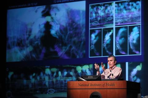
Photo: Chia-Chi Charlie Chang
Suddenly, the dynamics of the endoplasmic reticulum became visible. The fusions and fissions that characterize endoplasmic reticulum-mitochondria interactions became apparent. Myosin filaments could be seen in production and embryogenesis could be better studied. The tango of T cells and their antigens could be seen. Science’s ideas about how transcription worked were upended.
“I will never make a better contribution [to science] than lattice light-sheet microscopy,” Betzig said. “I never felt more like Galileo than with this microscope…Everyone who comes to us goes away with 10 terabytes (TB) of data and a big smile on their face.”
A ‘glass half-empty guy’
His collaborators are legion, numbering nearly 200, including many teams at NIH, where he and Hess worked in 2005. They did much of their work inventing PALM in a darkroom in Bldg. 32 with Dr. Jennifer Lippincott-Schwartz.
“If there’s one way I would describe the cell nowadays, and my understanding of it as a physicist,” said Betzig, “is it’s a highly stochastic [unpredictable] system...The smaller you look, the more stochastic it is. The dynamics is an incredibly important part of being able to understand how the cell works.”
He admitted, “Being the glass half-empty guy that I am, and the pessimist, I was disappointed with the lattice, despite its successes.” Most multi-cellular systems are like a bag of marbles or seeing through a wet windshield, because of their different refraction indices, said Betzig, who seems to work best when most perturbed.
He envisioned two paths to future improvements, both offered by his brethren who study the cosmos. “My take-home message is that microscopists are the retarded step-children of astronomers because we steal everything from them 50 years after they develop it first,” he quipped.
One “theft” is eliminating aberrations, such as those posed by the atmosphere for astronomers, who simply put their telescopes in orbit to solve the problem. In the past few years, biologists have used expansion microscopy to get rid of aberrations in their specimens. But that can’t be done on live animals.
The second path is adaptive optics, which Betzig likened to “a dynamic pair of eyeglasses.” His team combined AO technology with the lattice light sheet to study organelle dynamics in 3-D over time, even following cells through mitosis and watching zebrafish spinal cords grow from the inside out.
“I find biology beautiful, amazing and incredibly humbling and scary in the thought that there are 37 trillion cells in your body right now doing that dance,” he said, as a video of neutrophil motility in the zebrafish inner ear played on screen. “And each one of those cells has 2 billion molecules that are kind of doing the kind of dance you see in those SPT PALM movies…We really have our work cut out for us, to try to understand these systems. It’s amazingly complicated.”
As big a boon as new wave microscopy has been to biology, access to such expensive and rare tools is a barrier, Betzig noted. “The ultimate answer is commercialization” to remove the bottleneck.
Making a ‘Swiss Army knife’
He and his team have created what they call a “Swiss Army knife,” a next-generation lattice-AO microscope measuring 4 feet by 4 feet that can accommodate a range of sample sizes. Betzig said it incorporates, in one box, all of the last 30 years of improvements in microscopy. His colleagues at the Janelia Research Campus are building the first seven of them now, at a cost of about $410,000 a pop, and two more will form the cornerstones of the Advanced Bioimaging Center he is helping to establish at Berkeley.
Each knife will generate about 20 TB of data a week, which sounds great until you consider that data generated 5-6 years ago are just now making it into papers published today, Betzig said. Armies of skilled data analysts will be needed to parse a data tsunami that has already broken over biologists’ heads, he said, and big tech companies are raring to display their data-crunching chops.
The ability to see cellular dynamics “with clarity that we could not have imagined 20 years ago, 15 years ago…[is] going to change the way we look at living systems in the future,” Betzig concluded.
The complete talk is available at https://videocast.nih.gov/summary.asp?Live=31951&bhcp=1.
