NIH Record - National Institutes of Health
Cells from mouse skeletal muscle stem cells glow green.

July 9, 2021

  • Dr. Stephen Chanock

    Chanock Explores Genetics of Cancer Susceptibility

    At a recent virtual Clinical Center Grand Rounds Great Teachers talk, NCI's Dr. Stephen Chanock managed to fit his professional journey, career advice, a history lesson and decades of research into a captivating hour.
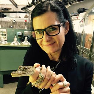
  • Johnson holds a baby crocodile in a lab

    DNA Forensics Help Solve Wildlife Crimes, Conserve Species

    Genomics can help solve animal cruelty cases, prosecute wildlife crimes and conserve endangered species, said Dr. Rebecca Johnson. “DNA can really be a valuable tool for fighting wildlife crime when combined with museum collections,” she said at the during the recent Louise M. Slaughter National DNA Day lecture.
  • Herr is smiling, seated at a table. His prosthetic legs are prominent. On a wall behind him is an artwork of a pair of legs.

    AMI Technique Gives New Life to Amputees

    In his recent WALS presentation, “On the Design of Bionic Limbs: The Science of Tissue-Synthetic Interface,” Dr. Hugh Herr spoke about a new surgical technique developed by his lab.
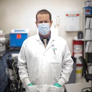
  • Dr. Mark Pandori

    Pandori Discusses What We Know About Covid Re-Infections

    Those who have recovered from SARS-CoV-2 may still be at risk for re-infection. Dr. Mark Pandori discusses efforts to identify re-infections and what these cases mean for the future of the pandemic.
Cells from mouse skeletal muscle stem cells glow green.

On the Cover

Multi-Nucleated Muscle Cells Grown in Culture. The cells have fused together to form myotubes that have many nuclei (stained blue). The cells are from mouse skeletal muscle stem cells treated with a harmless virus that caused them to glow green. The green color remained when the stem cells fused into myotubes. Some myotubes are stained red for a protein involved in muscle contraction (myosin heavy chain), a characteristic of mature muscle fibers. Researchers plan to use the same viral delivery system to genetically modify the cells and assess how impairing cell fusion alters myotube growth. The image was a 2017 winner in the BioArt competition of FASEB.

Photo: KEVIN A. MURACH, UNIVERSITY OF KENTUCKY, WITH SUPPORT FROM NIAMS, NIA

Back to Top