Out of Sight
NEI Scientists Publish Cell-Making Recipe for Research
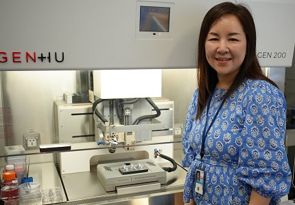
Photo: Dana Talesnik
Dr. Tea Soon Park and colleagues at the National Eye Institute (NEI) published a step-by-step protocol that uses human induced pluripotent stem cells (iPSCs) to produce key components of capillaries (tiny vessels that carry blood, oxygen and nutrients throughout the body). Now, they’re combining these capillary cell types to make tissues to study eye diseases and potential new approaches for treatments.
“Throughout the years, my research has been closely related to blood vessels and blood progenitor cells,” said Park, a scientist in NEI’s ocular and stem cell translational research section and lead author of the protocol. “During my postdoc, I studied the inner-retinal blood vessels using iPSC-derived regenerative therapy. Then came the idea of combining cell types to generate vascularized tissues using stem cell-derived cells.”
The protocol, published last year, details a system of how to produce three cell types necessary to generate capillary structure: endothelial cells—which line the capillaries; pericytes—which encase endothelial cells and stabilize the capillaries; and fibroblasts—which support and connect tissues. Park’s reproducible recipe takes the researcher through the steps to seed, differentiate and separate progenitor cells, and explains how to freeze and thaw them when ready for use.
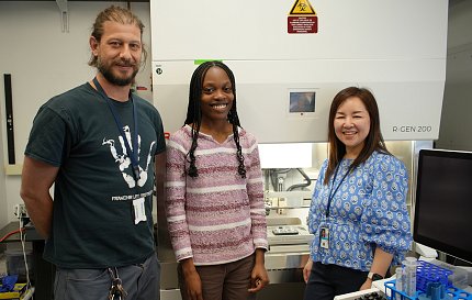
Photo: Dana Talesnik
Led by Drs. Kapil Bharti and Ruchi Sharma, the team is using this process to study tissue in the back of the eye, called the choroid. Dr. Andrea Barabino, a postdoctoral fellow in the lab and Casey Cargill, a postbaccalaureate fellow, are collaborating with Park on 3D-bioprinting technology to make vascularized choroid tissue [made from capillary cells].
The 3D-bioprinted choroid is combined with iPSC-derived functional ocular cells, called retinal pigmented epithelial (RPE) cells. These replicated, lab-made tissues allow researchers to study age-related macular degeneration (AMD) and other eye diseases in which capillary atrophy occurs during the disease progression.
“Our current project focuses on how the wet type of AMD begins, which occurs due to abnormal capillary growth under the macula,” Cargill said. AMD, which affects millions of elderly Americans, can lead to vision loss.
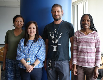
Photo: Dana Talesnik
AMD “is a disease that’s hard to mimic in animal models,” Barabino said, because only humans and primates have a macula. “That’s why we use human iPSCs to do in vitro modeling.”
Cargill explained, “We can use our iPSCs protocol to differentiate cells, make the choroid layer of the eye and mimic this disease.”
“One advantage of using iPSCs is we can target specific proteins or genes,” noted Park. “When studying AMD, we know the genetic risk factors so we can target those genes in specific cell types. With that, this protocol, will allow us to combine cells with genetic variants and investigate which types of cells are more damaged under stress conditions. That way we can trace back to where the degeneration of the retina is starting from.”
It’s an intricate system. “If any one cell type is not good in the system, we have to start all over again,” noted Devika Bose, an NEI stem-cell research associate who has regularly supplied members in OSCTR with iPSCs for the project.
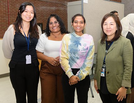
Photo: Marleen Van Den Neste
Another challenge, said Barabino, is that iPS cells can behave differently depending on their donor.
“It takes a lot of work to find the right concentration of all the molecules you want [in a way] that’s consistent across all cell lines,” he said. “Sometimes you have to change the parameters and tweak them to adapt to the new donor iPS cell line.”
Scientists from across the field developed this technology over many years. Bharti and Sharma’s group, though, is the first to publish a detailed recipe, based on prior research and their own, so others can reproduce the process.
The procedure also can be expanded beyond studying the eye, added Park. It can be used for any type of organ studies or condition involving the vascular system.
Photo: NEI
“This work requires close teamwork,” she emphasized. “It’s always helpful and inspirational to work together, especially on different parts of cells and specialized functions in the eye, because the eye is such a complex organ.”
“People in the industry are very interested in this 3D-bioprinted choroid/RPE tissue and want to know if it’s possible to make a product,” Park said. It might be possible to one day put the choroid and RPE together to make a treatment for late-stage AMD, she noted, but there is still a long way to go.
“I think that’s the future,” Bose said.
For more stories from NIH's Makers series, see: NIH Makers Series | NIH Record
