Going Down Memory Lane
NINDS Researchers Discuss the Mysteries of Memory
What—and how—do we remember?
This question is one of neuroscience’s many remaining mysteries. Memory is defined as the process of encoding, storing and retrieving information—a deceptively simple definition for a process that scientists are still trying to understand.
Researchers from the National Institute of Neurological Disorders and Stroke (NINDS)—Dr. Yi Gu and Dr. Kareem Zaghloul—spoke about their memory research at a recent Demystifying Medicine lecture series.
One way to understand it is to look at where it goes wrong. Patients who suffer from epilepsy may experience memory loss as a side effect of their seizures.
Zaghloul, a senior investigator and neurosurgeon in the NINDS Functional Neurosurgery lab, researches the activation of cortical networks during memory encoding and recall, and also works as a clinician. He treats patients with drug-resistant epilepsy by identifying and surgically removing the area of the brain where the patient’s seizures originate.
One contributor to epilepsy is scarring on the hippocampus, the region of the brain associated with memory and learning. Surgically removing the hippocampus would be much more likely to achieve remission than treatment with standard medications.
Zaghloul uses tools such as functional magnetic resonance imaging (fMRI) and electrodes on the patient’s scalp (electroencephalogram/EEG) to identify the “irritative zone” where the seizures originate. He can also surgically implant electrodes in the brain (intracranial EEG/iEEG) for more precise monitoring. Finally, once he has identified the source of the patient’s seizures, Zaghloul can surgically remove it.
How does this translate to memory research? Zaghloul’s patients who require an iEEG can be monitored in the hospital for up to two weeks while the device collects seizure data, and he has found they are often willing to participate in other, less invasive studies during that time. His research focuses on episodic memory—the memory of a specific event. By asking patients to play memory games and complete other related tasks, he can use the iEEG to observe episodic memory at work.
“We can ask what the pattern of activity looks like when you are forming a memory and, later on, when you are retrieving the memory, we see that you tend to repeat those same patterns,” Zaghloul said.
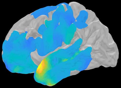
Photo: Zaghloul Lab, NINDS
He can have patients repeat these same tasks over a period of time and use that data to create a similarity map, which shows how similar the brain activity is across the iEEG electrodes as the patient tries to recall a memory.
“As the patient is trying to retrieve a memory and is about to say what they remember, they actually replay the same patterns from when they were encoding the memory,” said Zaghloul, confirming the trend he observed previously.
Since making this discovery, his lab has primarily focused its research on how this process actually occurs.
“What is underlying our ability to retrieve and relive memories?” Zaghloul asked. “How do we draw upon our memories to make decisions?”
Gu delved deeper into some of these questions in the second half of the lecture. Her lab studies the neural basis of spatial navigation and memory in rodents, essentially, what happens within the brain to let a rodent memorize and navigate its environment. This occurs in the medial temporal lobe, a structure that contains the hippocampus, the entorhinal cortex and several other elements that collaborate to form memories. Some of the cells within these regions have developed their own unique roles.
“Spatially modulated cells are everywhere in the entorhinal and hippocampal circuits,” Gu said, which underscores their importance in memory and navigation. She presented research on two specific cell types within the medial temporal lobe: place cells and grid cells.
Place cells are a type of neuron within the hippocampus that are active when an animal enters a specific place in its environment. Grid cells, which are found within the medial entorhinal cortex, are active at multiple places in an environment and these places are represented as vertices of a triangular lattice, forming a grid. These cell types likely work together to help animals form a “cognitive map” of their environment.

Gu introduced a study in which mice were trained to navigate in virtual reality (VR) to a “reward zone.” The mice licked the reward zone and received a treat. When the researchers recorded the activity in the rodents’ hippocampus, they could see the activation of place cells that represented the reward zone. If those place cells were stimulated when the mice were in a different area of the environment, they would lick at the area as if it were the reward zone, suggesting that place cell activity can directly drive behavior.
Gu also presented one of her studies on grid cell activity during learning. She first allowed mice to learn a VR environment until familiarization. Then she switched the mice to a new VR environment and studied their grid cell activity. They showed very different activity in the new environment, Gu reported, suggesting the grid cell activity “remapped” to encode information in the new environment. The similarity of grid cell activity continuously increased during learning, supporting its role in spatial memory.
Spatially modulated cells also seem to be affected in conditions such as Alzheimer’s disease, as Gu described. Researchers observed impaired place and grid cell activity in both a mouse model of the disease and in patients with Alzheimer’s when subjects were asked to complete a spatial memory task.
Using your medial temporal lobe may be a good defense against Alzheimer’s and memory decline, Gu said, citing a study showing that taxi and ambulance drivers experienced a lower incidence of memory impairment in comparison to drivers who followed a fixed route, such as bus drivers and pilots.
“The more you use your medial temporal lobe, the more protection you may have from disease and memory decline,” she concluded.
The moral of her talk? “Use your medial temporal lobe as much as you can!”
An archived version of the lecture can be viewed at go.nih.gov/6J3uLI8.