Serendipity, Symbiosis Strike
Basser Experiences Eureka Moment at NIH Research Festival

Dr. Peter Basser’s eldest brainchild is an invention that keeps on giving. Now after more than 33 years, new adaptations and applications of diffusion tensor magnetic resonance imaging (DTI) continue to spring forth from myriad scientists and disciplines. Conceived by chance at an NIH Research Festival, DTI serves as the poster child for the ingenuity of NIH’s Intramural Research Program.
“As a postdoc, I stumbled on a poster that my future colleague, [Dr.] Denis Le Bihan, and his colleagues in Radiology had presented at the 1991 NIH Research Festival…a venue for people in the Intramural Research Program to communicate ideas,” recalled Basser, NIH senior investigator and head of NICHD’s section on quantitative imaging and tissue sciences. “I wasn’t working on nuclear magnetic resonance [NMR] per se before that, although I had a patent issued from the late ‘80s on an NMR method. The invention of DTI was a happy accident. The work that I had been doing in the Biomedical Engineering and Instrumentation Program required high quality in vivo data—we had great models to describe important biophysical transport processes, but we needed better data to be able to inform these models.”
Now a principal investigator in NICHD’s Intramural Research Program, Basser uses noninvasive MRI methods to describe relationships between structure and function in living tissues—how their microstructure, hierarchical organization, composition and material properties affect the way they work…or don’t work.

In 1991, Basser had been at NIH for four years and had already established himself as a “serial inventor.” However, many models he and his group were producing leapt ahead of the technology to characterize them.
“Our biophysics-bioengineering models outstripped the methods that were available to provide parameters for them,” Basser recalled. “So I stumbled upon MRI as a methodology to fill this gap.”
He invented DTI after encountering Le Bihan’s group’s work while wandering through the back tent of Research Festival. It was serendipity.
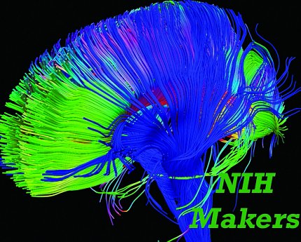
Diffusion tensor MRI addresses many problems. In neuroscience, for example—specifically neuroradiology clinical imaging applications—the concept can “better describe and understand the architecture of the brain and how different parts of the brain are connected…the gross wiring diagram of the brain,” Basser explained.
DTI also helps neurologists diagnose brain diseases and neurosurgeons navigate procedures.
“Neurosurgeons who are planning operations can use this information to avoid areas so that the patient doesn’t leave the surgery blind, or unable to hear or unable to understand language,” Basser said.
Radiation oncologists use the technology to kill tumors while sparing important brain areas that would cause disability. “They need to know what areas to avoid, that would leave the patient worse off,” Basser pointed out. Developmental biologists use DTI to understand how the brain forms and changes at different stages of life.”
Over the next several decades, Basser’s invention sprouted a number of other technologies that have become fields in themselves. “Tractography,” for example, allows scientists to discover how different areas of the brain are connected to one another, Basser added.

Photo: Peter Basser lab
“[DTI] is used to look at the structure of the heart now and not just inside the brain, but also in other tissues and organs in the body to see muscle architecture in the heart and tubule architecture in the kidney,” he noted. “It’s become a very sensitive indicator of tumor growth, not only in the brain, but in the rest of the body. It’s become built into a lot of clinical systems to diagnose cancer, and monitor therapy effectiveness. It’s used in a lot of specialties and subspecialties in medicine more and more.”
Basser and colleagues have continued to finetune DTI technology, even as dozens of applications for the method in various medical disciplines develop every few years. That it was born in-house on the Bethesda campus is a source of pride for intramural investigators in particular.
“I really feel fortunate that I was in the right place at the right time,” Basser concluded. “NIH has been a partner in all of this. Had we not had the Research Festival and had NIH not brought together these diverse groups of scientists, this invention never would have happened here.”
This story is part of an ongoing series on NIH Makers.
